


Dental diagnostics have advanced significantly with the integration of technologies like X-rays and intraoral camera examinations. X-rays offer high-precision insights into teeth, gums, and bones, aiding in detecting decay and infections. The intraoral camera examination provides non-invasive, high-definition visuals of 360 degrees inside the mouth, enhancing diagnostic accuracy and patient education on cosmetic and restorative treatments. This technology promotes patient engagement and active participation in maintaining optimal oral health, especially during routine check-ups.
In the realm of dental diagnostics, understanding the nuances of various examination tools is paramount for accurate health assessments. This article delves into two prominent methods: X-rays and intraoral camera examinations. We explore the intricacies of each technique, highlighting their respective advantages and limitations. By comparing these tools, we empower dental professionals and patients alike to make informed decisions, fostering better oral healthcare management through detailed visual insights provided by both X-rays and intraoral cameras.
- Understanding X-rays: What They Reveal About Dental Health
- The Role of an Intraoral Camera: Capturing Detailed Visuals
- Comparing Methods: Advantages and Limitations of Each Examination Tool
Understanding X-rays: What They Reveal About Dental Health
X-rays have long been a cornerstone of dental diagnostics, offering valuable insights into the intricate world of oral health. These advanced imaging tools allow dentists to peer beyond what the naked eye can see, revealing critical information about teeth, gums, and bone structures. When it comes to detecting decay, infections, or abnormalities, X-rays are indispensable. They can pinpoint areas of dental caries, track the progress of periodontal disease, and even expose hidden cysts or tumours. By providing detailed, high-resolution images, X-ray examinations offer dentists a clear roadmap for developing effective treatment plans, encompassing both preventative measures and restorative procedures.
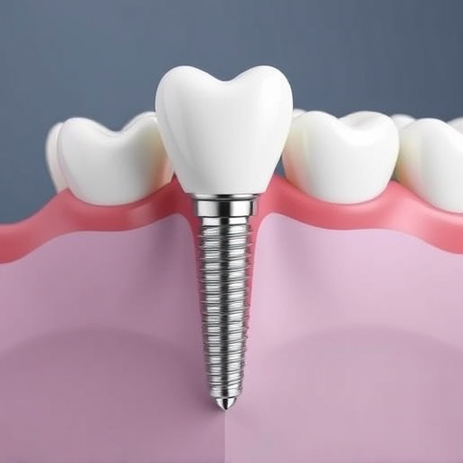
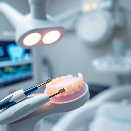
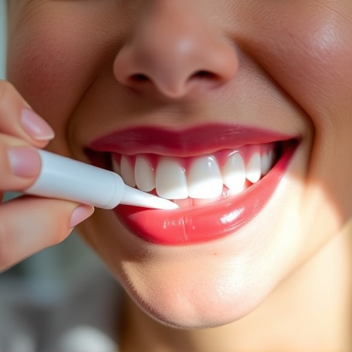
Complementing traditional X-rays is the intraoral camera examination, which brings a new dimension to dental assessment. This non-invasive technology captures visual data at a microscopic level, allowing patients to see their oral health from a dentist’s perspective. By integrating real-time images into regular check-ups, dentists can more accurately diagnose issues and educate patients about cosmetic dentistry or restorative treatments, such as dental implants. The intraoral camera examination facilitates patient engagement, empowering individuals to actively participate in maintaining optimal dental health.
The Role of an Intraoral Camera: Capturing Detailed Visuals
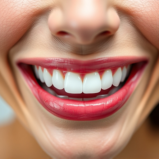
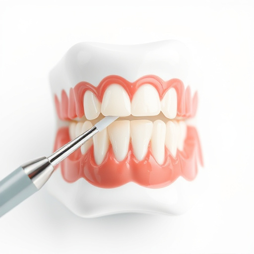
An intraoral camera plays a pivotal role in modern dental examinations, revolutionizing how dentists assess and diagnose oral health issues. This advanced technology allows for the capture of detailed, high-resolution visuals of teeth, gums, and the oral cavity. By integrating an intraoral camera examination into routine oral exams, dentists gain a clear and comprehensive view of areas traditionally hard to inspect, such as tooth surfaces, fillings, and bonding.
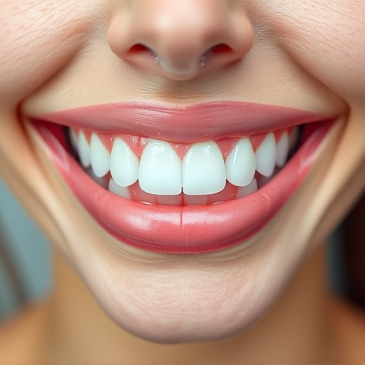
Compared to traditional X-rays, which offer valuable insights but are 2D projections, the intraoral camera provides a 360-degree, visual perspective in high definition. This detailed visualization enables dentists to more accurately identify decay, cracks, or other abnormalities that might be missed by conventional methods. Moreover, it aids in patient education during routine dental check-ups, allowing patients to better understand their oral health and the condition of their dental fillings or bonding.
Comparing Methods: Advantages and Limitations of Each Examination Tool
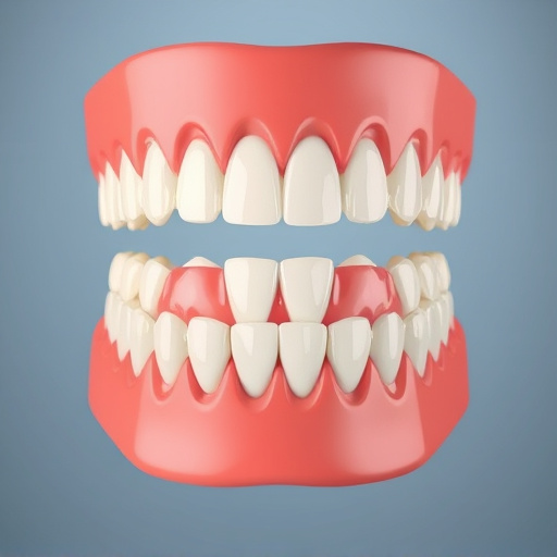
Comparing X-rays and intraoral camera examinations offers valuable insights into dental health. Each method has its unique advantages and limitations. X-rays, being a traditional tool, provide detailed images of dental structures, including bones and teeth, making them indispensable for diagnosing conditions like cavities, infections, and bone loss. They are particularly crucial in planning treatments such as tooth extractions and cosmetic dentistry procedures like dental bonding.
Intraoral cameras, on the other hand, offer visual accuracy and a non-invasive approach. These high-resolution cameras capture detailed images of teeth and gums from within the mouth, enabling dentists to identify issues like early tooth decay, gum disease, or even abnormalities in dental alignment. This technology is particularly beneficial for cosmetic dentistry patients as it allows precise evaluation of the oral cavity before and after procedures, such as bonding or other aesthetic modifications. However, intraoral cameras may not detect certain conditions that are better visualized with X-rays.
In comparing X-rays and intraoral camera examinations, both offer valuable insights into dental health. X-rays provide detailed images of bone structure and hidden areas, while intraoral cameras capture high-resolution visuals of teeth and gums. Each method has its advantages and limitations; X-rays are essential for diagnosing conditions like cavities and bone loss, whereas intraoral cameras enable more visual assessments of surface issues and gum health. Combining these tools offers a comprehensive understanding of oral health, ensuring better patient care and treatment planning.